
Book Appointment Now

Mammography
High-precision, low-dose breast imaging for early detection of lumps, microcalcifications, and tissue changes.
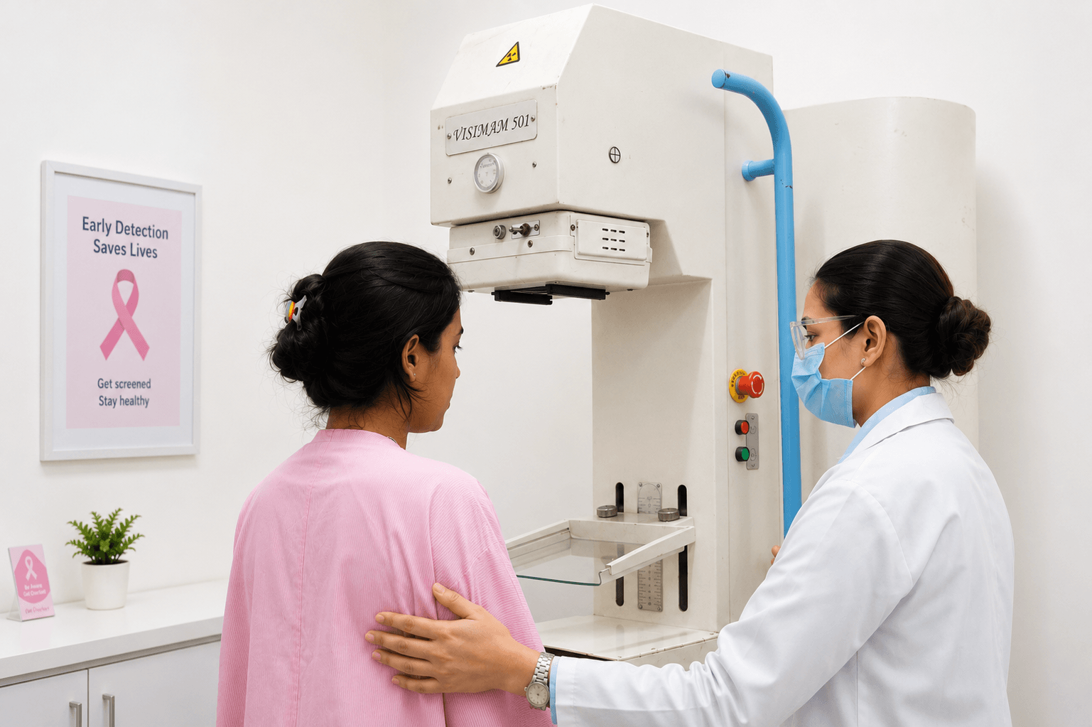
Digital Mammography & 3D Breast Tomosynthesis
It is the single most effective tool worldwide for detecting early-stage breast cancer up to 3 years before a lump can be physically felt.
Commonly Booked Tests
Prerequisite:
KFT / Serum Creatinine Report Required
KFT / Serum Creatinine Report Required
Scan Duration:
10–15 Minutes
10–15 Minutes
Preparation:
Avoid deodorants, talcum powders, or lotions on scan day
Avoid deodorants, talcum powders, or lotions on scan day
Report Delivery:
Within 1 Hours
Within 1 Hours
What to expect
You will be guided by an experienced female mammography technologist in a private, gentle environment.
Each breast is briefly compressed between two smooth clear plates for 5 to 10 seconds to flatten tissue and optimize image detail.
Each breast is briefly compressed between two smooth clear plates for 5 to 10 seconds to flatten tissue and optimize image detail.
Prep Instructions
Do not apply deodorants, antiperspirants, powders, ointments, or creams under your arms or on your breasts. Metallic particles in these products can mimic calcifications on the scan.
Wear a top and bottom so you only need to undress above the waist.
Timing Tip: Schedule your scan for the week after your menstrual cycle when breasts are least tender.
Wear a top and bottom so you only need to undress above the waist.
Timing Tip: Schedule your scan for the week after your menstrual cycle when breasts are least tender.
FAQ’s
Why are deodorants and powders forbidden on the day of a mammogram?
Many deodorants, powders, and lotions contain small metallic particles (like aluminum) or zinc. On a digital mammogram, these specks can appear as bright white spots that look identical to microcalcifications (early warning signs of tissue changes), leading to unnecessary re-scans.
Is mammography compression painful?
Compression may cause brief discomfort or pressure for a few seconds per view, but it should not be painful. Firm compression is necessary to spread out breast tissue, reduce radiation dose, and prevent motion blur.
What is the difference between 2D Digital Mammography and 3D Tomosynthesis?
A standard 2D mammogram takes one flat image from the front and side. 3D Tomosynthesis captures multiple thin-slice images at different angles, allowing the radiologist to look through dense breast tissue layer-by-layer.