Book Appointment Now

Colour Doppler
Advanced 2D/3D non-invasive acoustic imaging to evaluate real-time blood flow, arterial blockages, deep vein thrombosis (DVT), and fetal circulation.
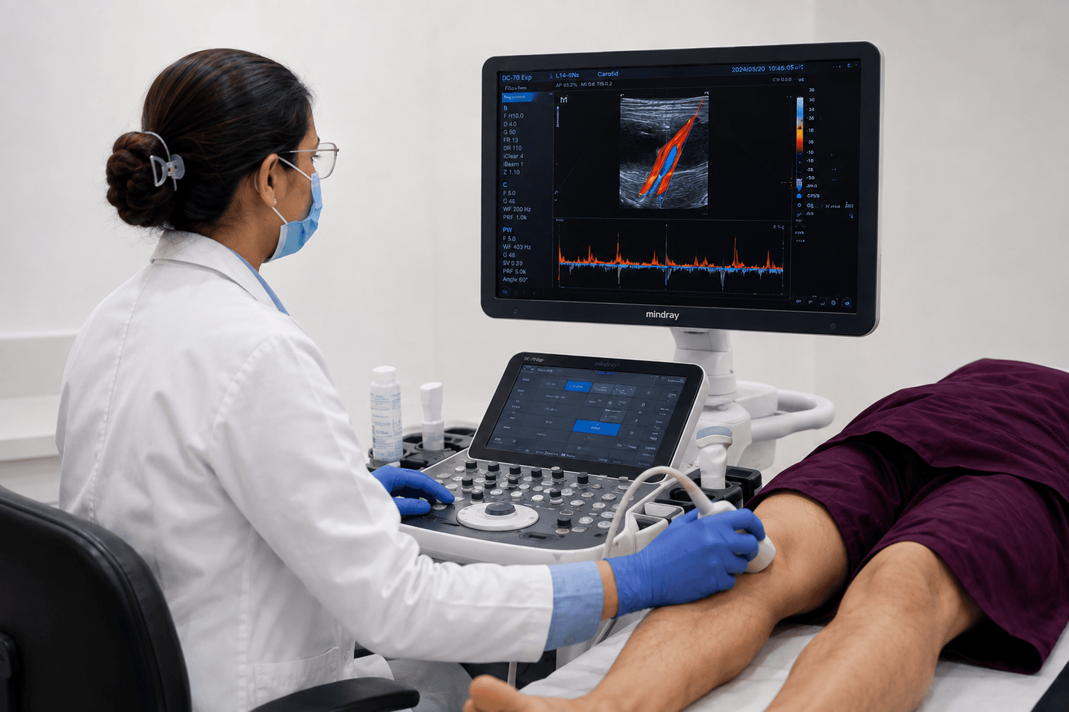
Color Doppler Ultrasound
An ultrasound-based scan that studies blood flow through arteries and veins, helping detect blockages, clots and circulation issues — commonly used for leg pain, varicose veins, and pregnancy blood-flow checks.
Unlike standard grayscale ultrasound—which shows static organ structures—Color Doppler converts sound waves reflected off moving red blood cells into vivid red and blue color maps on screen. This allows senior radiologists to assess the speed, direction, and turbulence of blood flow through your vessels without needles, radiation, or contrast dyes.
Unlike standard grayscale ultrasound—which shows static organ structures—Color Doppler converts sound waves reflected off moving red blood cells into vivid red and blue color maps on screen. This allows senior radiologists to assess the speed, direction, and turbulence of blood flow through your vessels without needles, radiation, or contrast dyes.
100% Safe:
Zero ionizing radiation or harmful rays
Zero ionizing radiation or harmful rays
Scan Duration:
15–30 Minutes
15–30 Minutes
Preparation:
Fasting or full bladder depending on site
Fasting or full bladder depending on site
Report Delivery:
Within 1 Hours
Within 1 Hours
Commonly Booked Tests
What to expect
You will lie comfortably on an examination couch.
Warm, water-based acoustic gel is applied to the skin over the target area.
The radiologist moves a hand-held transducer (probe) across the skin. You will hear swooshing audio sounds (pulse-wave signals) representing real-time blood flow.
Warm, water-based acoustic gel is applied to the skin over the target area.
The radiologist moves a hand-held transducer (probe) across the skin. You will hear swooshing audio sounds (pulse-wave signals) representing real-time blood flow.
Prep Instructions
Abdominal & Renal Doppler: Fast strictly for 4 to 6 hours before your appointment to minimize bowel gas.
Obstetric / Fetal Doppler: Drink 2–3 glasses of water 1 hour before the scan (a comfortably full bladder may be required).
Limb / Carotid Doppler: No fasting or special diet required. Wear loose, comfortable clothing.
Obstetric / Fetal Doppler: Drink 2–3 glasses of water 1 hour before the scan (a comfortably full bladder may be required).
Limb / Carotid Doppler: No fasting or special diet required. Wear loose, comfortable clothing.
FAQ’s
Is Color Doppler safe during pregnancy?
Yes, absolutely. Color Doppler relies purely on safe, high-frequency sound waves (ultrasound) and uses zero ionizing radiation. It is widely prescribed during high-risk pregnancies to monitor placental nutrition and fetal blood supply safely.
Is Doppler different from a regular ultrasound? What do the red and blue colors on the Doppler screen mean?
It uses the same technology as ultrasound but specifically maps blood flow and speed, in addition to structural imaging. The colors represent the direction of blood flow relative to the ultrasound probe, not whether the blood is oxygenated or deoxygenated. By standard convention:
Blue: Blood flowing away from the transducer probe.
Red: Blood flowing towards the transducer probe.
How does Color Doppler detect a Deep Vein Thrombosis (DVT)?
The radiologist uses compression ultrasound and Color Doppler to examine deep leg veins. If a blood clot (DVT) is present, the vein will fail to compress under gentle probe pressure, and the color signal will show a complete or partial absence of blood flow around the clot.
Why do I need to fast before a Renal or Abdominal Doppler?
Fasting for 4 to 6 hours reduces gas in your stomach and intestines. Bowel gas scatters sound waves and blocks the ultrasound beam from reaching deep retroperitoneal vessels like the renal arteries and abdominal aorta.